Thanks to radiology signs
See the caseTrans-scaphoid-perilunate-dislocation


Disorder
|
Vasculitis
|
Epidemiology/Etiology
|
Clinical/Laboratory Findings
|
Takayasu's arteritis ("pulseless
disease")
|
Granulomatous large vessel vasculitis
involving aortic arch vessels
|
Young Asian women and children
|
Absent upper extremity pulse
Visual defects, stroke |

Disorder
|
Vasculitis
|
Epidemiology/Etiology
|
Clinical/Laboratory Findings
|
Giant cell (temporal) arteritis
|
Granulomatous large vessel vasculitis
involving superficial temporal and ophthalmic arteries; thrombi contain
microabscesses
|
Adults > 50 years of age
|
Temporal headache, jaw claudication
(pain when chewing)
Blindness on ipsilateral side Polymyalgia rheumatica (muscle and joint pain; normal serum creatine kinase) Increased ESR |


Disorder
|
Vasculitis
|
Epidemiology/Etiology
|
Clinical/Laboratory Findings
|
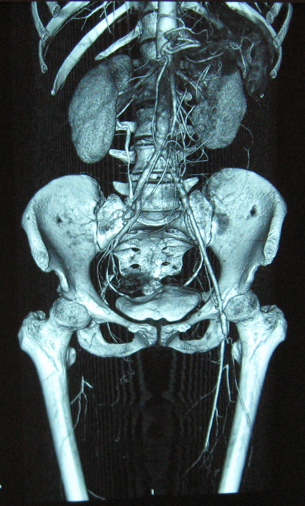
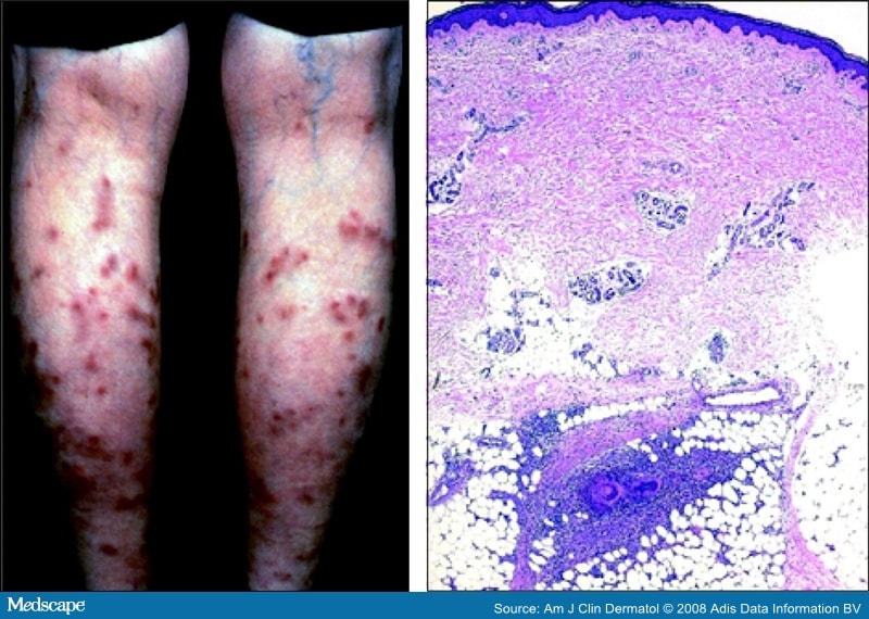
Polyarteritis nodosa
|
Necrotizing medium-sized vessel
vasculitis involving renal, coronary, mesenteric arteries (spares pulmonary
arteries)
|
Middle-aged men
Association with HBsAg (30%) |
Vessels at all stages of acute and
chronic inflammation
Focal vasculitis produces aneurysms (detected with angiography) Organ infarction in kidneys (renal failure), heart (acute MI), bowels (bloody diarrhea), skin (ischemic ulcer) |
Disorder
|
Vasculitis
|
Epidemiology/Etiology
|
Clinical/Laboratory Findings
|
Kawasaki disease
|
Necrotizing medium-sized vessel
vasculitis involving coronary arteries (e.g., thrombosis, aneurysms)
|
Children < 4 years of age
|
Desquamating rash, swelling of hands
and feet, cervical adenopathy, oral erythema
Abnormal ECG (e.g., acute MI) Corticosteroids contraindicated (danger of vessel rupture) |

Disorder
|
Vasculitis
|
Epidemiology/Etiology
|
Clinical/Laboratory Findings
|
Thromboangiitis obliterans (Buerger's
disease)
|
Medium-sized vessel vasculitis with
digital vessel thrombosis
|
Men 25-50 years of age who smoke
cigarettes
|
Foot claudication, Raynaud's
phenomenon, ulceration, gangrene
|